


Overview
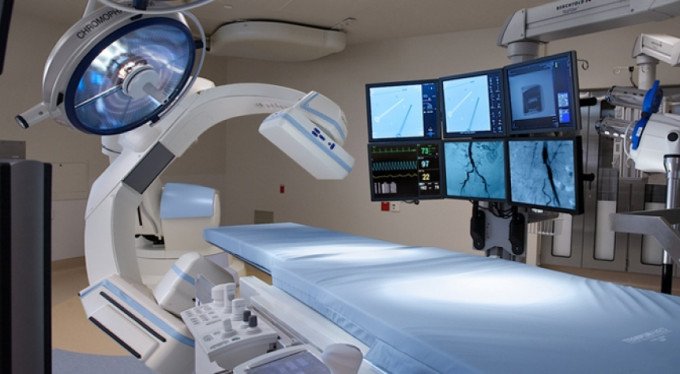
Interventional Radiology: interventional radiology implies diagnostic or therapeutic interventional procedures using imaging devices, such as US, CT, MRI or fluoroscopy. These devices visualize organs and tissues in procedures and thus, procedure-related risks are minimized. Procedures carried out in interventional radiology unit are addressed in two categories; vascular and non-vascular. Vascular procedures cover arteries and veins, while non-vascular procedures visualize all organs.
Vascular Interventional Radiology Procedures; vascular procedures are related to arteries and veins. For diagnostic procedures, special needles and catheters are inserted into the intended blood vessel and images are acquired by instilling tracers or contrast agents. Therapeutic procedures intend to solve problems that develop in blood vessels. A local anesthetic agent is administered and needle is inserted into an artery at the location of groin; next, a small-bore sheath is placed and a thin catheter is advanced inside this sheath to access the blood vessels and images are acquired, after contrast agent is instilled. Blood vessels are visualized and pathological conditions are revealed out after contrast agent is administered.
Diagnostic Vascular Radiology Interventions performed in our Unit:
4-system selective cerebral angiography with femoral puncture: it aims visualization of cerebral blood vessels.
Bilateral carotid angiography: visualizes arteries of the neck, called carotid artery. There are two carotid arteries in the neck, one at each side of the neck.
Selective vertebral angiography: visualizes vertebral artery in the neck. There are two vertebral arteries in the neck, one at each side of the neck.
Aortography: visualizes the aorta – the greatest artery that originates from the heart.
Aortofemoropopliteal (AFP) angiography: visualizes blood vessels of the brain.
Bronchial arteriography: visualizes the pulmonary arteries that flow blood into lungs.
Celiac angiography and arterial portography: visualizes the blood vessels that extend to liver and spleen.
Superior and inferior mesenteric angiography: visualizes blood essels of small intestines and colon.
Renal angiography: visualizes arteries of kidneys.
Pelvic angiography: visualizes blood vessels in lower part of the body.
Spinal angiographic screening: locates and visualizes arteries that feed the spinal cord.
Brachial angiography: visualizes arteries of arms.
Transplant renal angiography: it aims visualizing arteries and veins of kidneys in kidney transplant patients.
Inferior and superior venacavagraphy: visualizes major veins that flow blood into the heart.
Upper Extremity venography: visualizes veins of arms.
Renal venography: visualizes veins of kidneys.
Renal – petrosal blood sampling: blood specimens are drawn from veins, where hormones of adrenal glands and pituitary gland are secreted, and specimens are analyzed.
Hepatic venography: visualizes veins of liver.
Splenoportography. Visualizes veins of spleen and the portal vein that drains into the liver.
Testicular venography: visualizes veins of testicles.
Lower Extremity venography: visualizes veins of legs.
Arteriovenous fistulography: evaluates AV fistula in dialysis patients with AV fistula.
Evaluating patency of central venous catheter: catheter is evaluated in catheterized patients.
Therapeutic Vascular Radiology Interventions performed in our Unit:
Placement of vena cava filter: migration of clot from veins of legs to blood vessels of lungs is an extremely fatal condition. These filters hinder migration of these clots to lungs. A vein of groin or neck is punctured after local anesthetic agent is administered; a filter is inserted into the vena cava that flows blood from legs to the heart immediately beneath the renal veins and thus, clots cannot reach lungs. These filters are divided into two categories; temporary and permanent. Temporary filters are removed within 1 to 2 weeks, while permanent filters stay in the body for lifetime.
Tumor embolization: certain tumors can be embolized through procedures that directly manipulate the tumor or the feeder artery of the tumor.
Chemoembolization: chemotherapeutic agents are administered into the artery that feeds the tumor, especially in liver tumors.
Percutaneous transluminal angioplasty (PTA): a very thin wire is advanced through the stenotic segment that is detected in angiography and a balloon is placed and inflated at the stenotic part of the blood vessel. Thus, stenosis is eliminated and blood flow is improved.
PTA – Placement of stent: angioplasty cannot help stenosis or it may relapse. In such cases, wire cages are placed to the location of stenosis followed by balloon dilatation to expand the stent to the diameter of the blood vessel. Thus, patency is achieved and the blood flow is restored to the intended level.
Selective thrombolytic therapy: clot formation in lumen of blood vessels stops or impairs blood flow. Catheter is advanced into lumen of the diseased blood vessel and thrombolytic agents (drugs that lyse clots) are administered and thus, blood flow can be restored.
Tunneled catheter insertion: permanent and tunneled catheters are inserted into great arteries or central veins in patients who require dialysis therapy, but do not have an access; these catheters enable these patients have dialysis therapy. Useful life of these catheters is approximately 1 year.
Temporary catheter insertion: a temporary catheter is inserted into a central vein of the body for patients who require short-term dialysis or intravenous nutrition. Useful life of these catheters is approximately 15 days.
Subcutaneous port insertion: administration of chemotherapeutic agents into veins of cancer patients, who require chemotherapy, cases obstruction of the punctured veins. Therefore, chemotherapeutic agents are administered through ports. The port material is usually placed in subcutaneous tissue usually at upper part of the chest wall under local anesthesia and the port catheter is extended into the central vein that drains into the heart.
Removal of Tunneled Catheter: the previously inserted catheter is removed under local anesthesia.
Removal of Subcutaneous Port: the previously inserted port material is removed under local anesthesia.
Peripheral atherectomy – thrombectomy or laser: calcified plaques in feeder arteries of legs are removed using special devices and thus, blood flow is restored.
Laser or sclerosing agent therapy for varicose veins: the previously inserted permanent catheter is removed under local anesthesia.
Non-vascular Interventional Radiology Procedures
Non-vascular procedures intend non-vascular body structures. The procedures that intend body organs were carried out according to their anatomic position, before imaging modalities were developed. For instance, if a body organ, such as liver or kidney, would be biopsied, a specimen was taken by puncturing an anatomic zone that was corresponding to the organ without any visualization. In such cases, the physician could not see the organ to be biopsied or what lies in front of the targeted organ. If an organ is biopsied without imaging guidance, the risks include bleeding or taking specimen from inappropriate location. Now, interventional radiologists see the organ to be biopsied and its periphery using imaging devices and thus, the blood vessels and nerves inside or at the periphery of the organ are not damaged. For example, the thyroid gland is visualized under ultrasound guidance for thyroid biopsies and the thyroid nodule is accessed through the shortest route and precisely aspirated.
Therapeutic Vascular Radiology Interventions performed in our Unit:
- Biopsies: local anesthetic agent is administered and small tissue pieces are taken from body organs or tissues, such as thyroid, liver, kidney and lung.
- Ultrasound-guided breast marking: if the surgery aims isolated removal of the pathologic formation in the breast, interventional radiologist places a wire into the tissue, while one end of the wire is exposed on the skin; thus, access of surgeons to the tissue is facilitated.
- Paracentesis/Thoracentesis: a sample is drawn from the fluid located in pleura or membranes that cover abdominal organs.
- Percutaneous drainage of ascites/pleural effusion: a sample is drawn from the fluid located in pleura or membranes that cover abdominal organs.
- Percutaneous drainage of abscess/empyema: a catheter is used to drain an abscess located at any body part or the infected fluid located in pleura.
- Percutaneous treatment of cyst: a catheter is inserted to administer certain substances that can treat enlarged renal cysts, which cause clinical signs.
- Percutaneous treatment of pneumothorax: drainage of the air entrapped in pleura.
- Percutaneous drainage of lymphocele: the lymph that accumulates in any part of the body is drained.
- Percutaneous treatment of pseudocyst: a catheter is inserted to treat a certain type of cyst, called pseudocyst that develops following pancreatitis.
- Percutaneous treatment of hydatid cyst: a specific cystic lesion of liver, called hydatid cyst, is treated using catheters.
- Cholangiography: visualizes the bile ducts.
- Percutaneous transhepatic cholangiography: a thin needle is inserted into the biliary tree that is visualized after contrast agent is administered.
- Percutaneous drainage of biliary tree: a catheter is inserted into the dilated bile ducts to maintain bile flow.
- Percutaneous balloon dilatation of choledochus: a balloon is inserted and dilated to eliminate obstruction or stenosis of the common bile duct, called choledochus.
- Percutaneous biliary stent placement: a stent is inserted into the common bile duct to ensure the patency.
- Percutaneous drainage of gall bladder: a catheter is inserted and the expanded gall bladder is drained and emptied.
- Percutaneous alcohol ablation: a catheter is inserted into arteries of the kidney that is small and dysfunctional and causes hemodynamic disorders and alcohol is instilled to stop functioning of the kidney completely.
- Antegrade pyelography: a thin needle is inserted into collecting system of the kidney and contrast agent is instilled to acquire images.
- Ultrasound-guided percutaneous nephrostomy: an ultrasound-guided drainage catheter is inserted into the collecting system of kidney.
- Percutaneous dilatation of ureteral stenosis: the ureteral stenosis is dilated using a balloon.
- Percutaneous placement of ureteral stent: a stent is placed at the stenotic segment of the ureter to achieve patency.
- Percutaneous cystostomy: a catheter is inserted into dilated bladder that causes excretion problems in order to drain the urine.
- Percutaneous puncture of renal cyst: Renal cysts are drained using a needle or catheter and a sclerosing agent is injected into the cyst cavity.
- Pouch graphy: the ureteral stenosis is dilated using a balloon.
- Percutaneous gastrostomy: a catheter is inserted into lumen of stomach through the abdominal skin for patients who cannot be fed by mouth. Thus, nutritional products can be given through the catheter.
- Percutaneous gastrojejunostomy: a catheter is inserted into lumen of stomach and small intestine through the abdominal skin for patients who cannot be fed by mouth.
- CT-guided interventional examinations: guidance of computerized tomography is used for interventional procedures for patients, if quality of sonographic imaging is poor due to patient factors.
Doctors